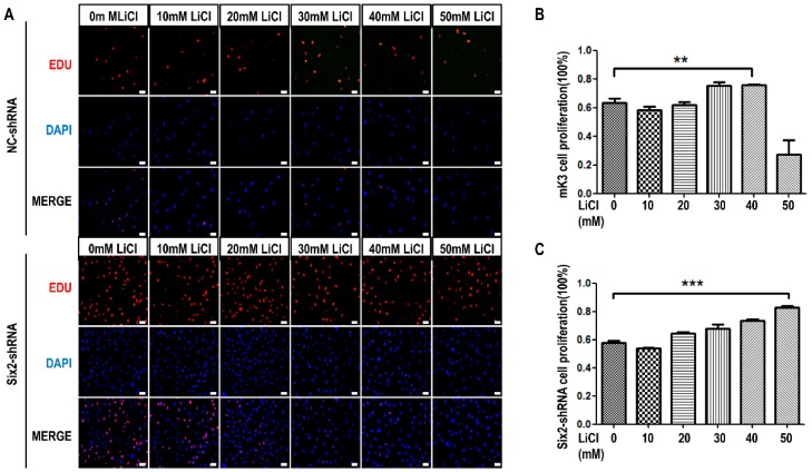
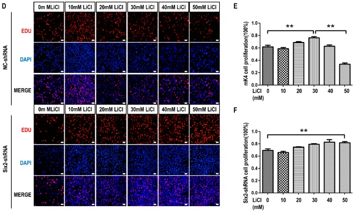
Figure 4.
Knockdown of Six2 gene inhibits cell proliferation while LiCl treatment of low-concentration promotes cell proliferation in mK3 and mK4 cells. (A) mK3 cells were transfected with negative shRNA control and Six2-shRNA for 36 h and treated with LiCl of increasing dosages for 12 h. Proliferating mK3 cells were labeled with EdU (red) and cell nucleus were stained with hoechst (blue). The EdU results were accessed by fluorescent microscope (200×) with the scale bar representing 20 μm and the respective pictures were merged to the purple one; (B,C) Statistical analysis of cell proliferation. Values were presented as mean ± SEM (n = 3), ** p < 0.01, *** p < 0.001 relative to control; (D) mK4 cells were transfected and were detected by EdU assay as same as mK3 cells in (A); (E,F) Statistical analysis of cell proliferation. Values were presented as mean ± SEM (n = 3), ** p < 0.01 relative to control.


