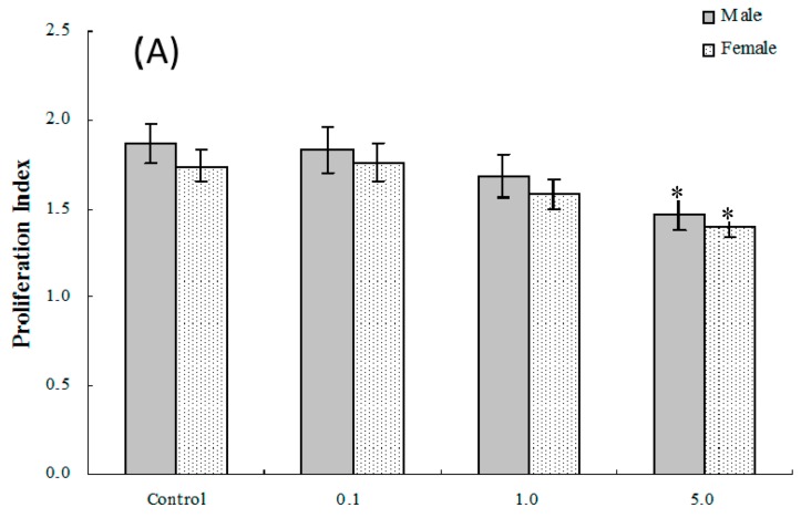
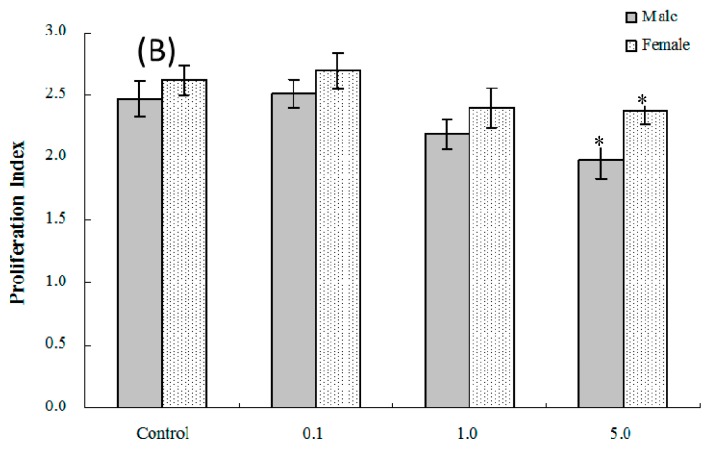
Figure 2.
Ex vivo splenic lymphocyte proliferation among splenocytes isolated from in C57BL/6 pups exposed in utero to PFOS during GD 1–17. (A) T and (B) B cell responses. Splenocytes from four-week-old hosts were isolated and then cultured (5 × 10⁶/well) with or without 10 μg/mL concanavalin A (ConA) or LPS for 48 h, before proliferation was measured using MTT. A value of 1.0 for proliferative index = proliferation obtained for non-treated (stimulated) cells. Unstimulated counts were not significantly different between groups. Data shown are the mean ± SEM. * Value significantly different from the control (p ≤ 0.05) by the one-way ANOVA test. n = 12 each group.


