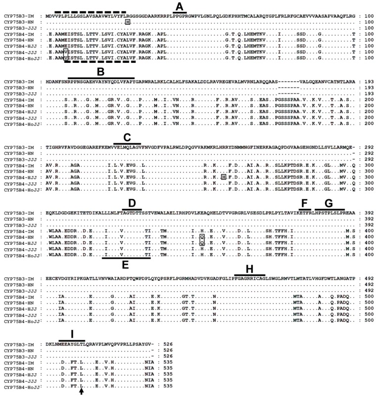
Figure 2.
Amino acid sequence alignment of CYP75B3s and CYP75B4s. Deduced amino acid sequences of CYP75B3 and CYP75B4 proteins from the white (IM), black (HN and HJJ), and red rice varieties (JJJ and HoJJ) were compared. Amino acid substitutions were indicated with rectangles. The two dashed lines located in the N-terminal region indicate the predicted membrane spanning anchors of CYP75B3 and CYP75B4 and the solid lines indicate specific conserved regions of cytochrome P450 enzymes. A: hinge region, B: substrate recognition site 1 (SRS1), C: SRS2, D: oxygen binding pocket, E: SRS4, F: ExxR motif, G: SRS5, H: heme biding domain, and I: SRS6. Thr504 of CYP75B3 and Leu512 of CYP75B4 correspond to the previously described functional determinant for F3′H activity, which are indicated by an arrow.

