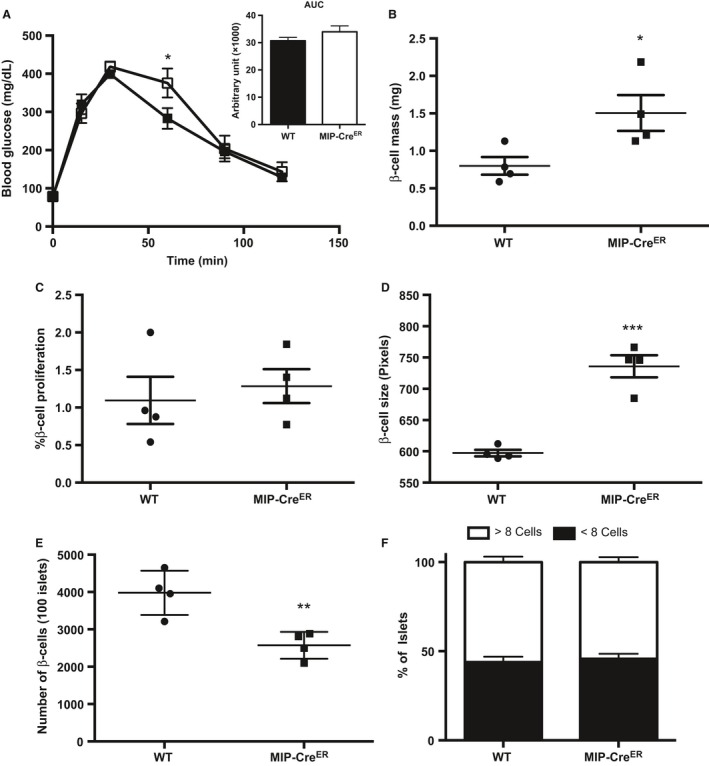
Figure 2.

Wild‐type (WT) and MIP‐CreER mice show similar levels of β‐cell proliferation in response to 1 week high fat diet (HFD) feeding. (A) Intraperitoneal glucose tolerance tests (IP‐GTT) and area under the curve (AUC) (WT n = 6, MIP‐CreER n = 3), (B) β‐cell mass, (C) β‐cell proliferation, (D) β‐cell size, (E) β‐cell number, and (F) percentage of small insulin+ clusters for 8‐week‐old WT or MIP‐CreER male mice placed on a HFD for 1 week. Black boxes represent WT mice; open boxes represent MIP‐CreER mice. Data are shown as means ± SEM. P values were calculated using Student's t‐test for (B–D) and two‐way ANOVA followed by Tukey post hoc analysis for (A). *P < 0.05 in (A); *P = 0.0385 in (B); and ***P = 0.0003 in (D); **P = 0.0067 in (E).
