Figure 3.

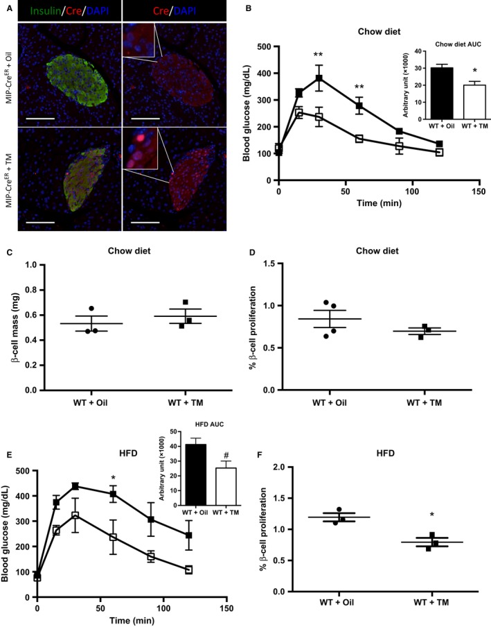
Tamoxifen (TM) inhibits β‐cell proliferation after 3 days high fat diet (HFD) feeding in Wild‐type (WT) mice. (A) Immunolabeling of Cre localization in MIP‐CreER mice injected with corn oil (top panels) or TM (bottom panels). In the absence of TM, Cre displays cytoplasmic localization (top panels). Upon TM treatment, Cre immunolabeling can be visualized in the nuclei, as marked by DAPI (bottom panels). Scale bars represent 100 μm. (B) Intraperitoneal glucose tolerance tests (IP‐GTT) and area under the curve (AUC) (WT + oil n = 3, WT + TM n = 3), (C) β‐cell mass, and (D) β‐cell proliferation for chow‐fed WT mice injected with corn oil or TM at 6 weeks of age and analyzed at 8 weeks of age. (E) IP‐GTT and AUC (WT + oil n = 3, WT + TM n = 3) and (F) β‐cell proliferation for WT mice placed on HFD for 3 days following the washout period. Black boxes represent WT + Oil‐treated mice; open boxes represent WT + TM mice. Data are shown as means ± SEM. P values were calculated using Student's t‐test for (C), (D), and (F) and two‐way ANOVA followed by Tukey post hoc analysis for (B) and (E). *P = 0.0291, **P < 0.01 in (B); # P = 0.0696, *P < 0.05 in (E); and *P = 0.0133 in (F).
