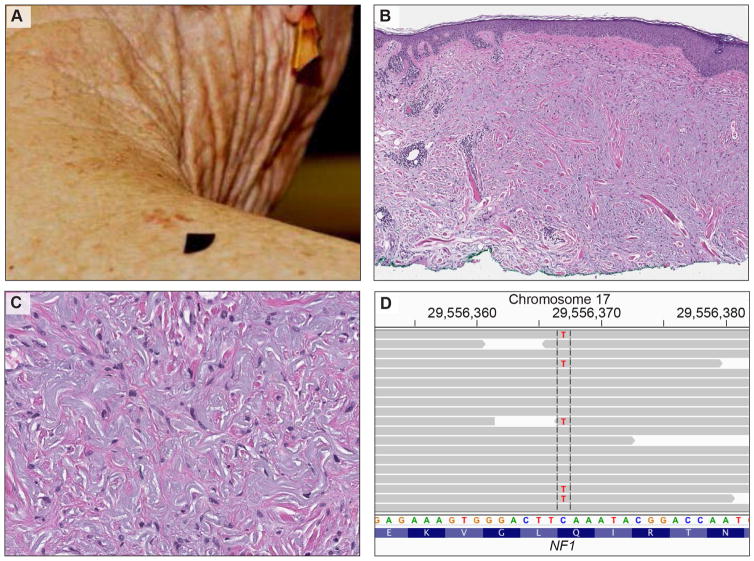
Figure 1.
Clinical, histological, and genetic features of desmoplastic melanoma. A, clinical picture of an 81-year-old woman with an irregular, brown-reddish skin lesion on her right shoulder. B, Histopathology of desmoplastic melanoma: Melanoma in situ (atypical proliferation of solitary units and nests of melanocytes at the dermoepidermal junction) is associated with an infiltrative pauci-cellular fibrosing spindle cell proliferation in the dermis with focal lymphocytic aggregates. C, The dermal spindle cells are dispersed as individual units (not aggregated in sheets or fascicles). They have hyperchromatic nuclei. D, The targeted sequencing data displayed in the Integrative Genomics Viewer (IGV). The gray arrows illustrate the individual sequencing reads aligned to exon 21 of the NF1 gene. The cytosine at base pair position 2734 was replaced by thymine (c.2734C>T) leading to a premature stopcodon (p.Q912*) and to a truncated NF1 protein.

