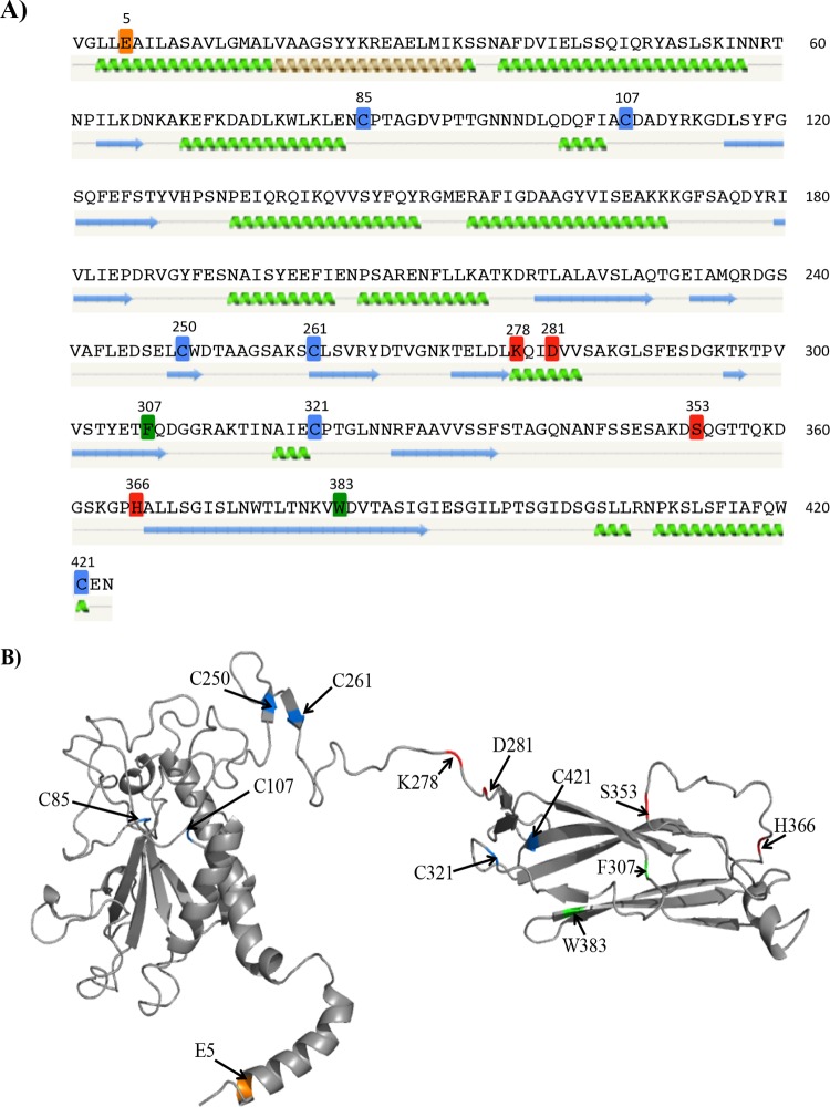
FIG 5.
Selected residues mapped onto predicted TcpB structural models. Residues that do not affect the stability of TcpB but impact the phenotypic profile of TCP, as well as the cysteine residues, are mapped onto the predicted secondary structure (A) and tertiary structure (B). (A) Cysteines (blue), E5 (orange), residues that primarily affect TcpF secretion (red), and residues that have an intermediate effect (green) are highlighted on the mature TcpB amino acid sequence. The predicted secondary structure of the protein generated using Phyre2 (33) is shown below the amino acid sequence. Green coils represent α-helices, blue arrows represent β-sheets, and gray lines indicate coils. (B) Theoretical structural cartoon model of TcpB created using Phyre2. The predicted structure was modeled largely on the crystal structure of CofB; however, the N-terminal region was modeled based on the full-length PAK pilin. Residues of interest are highlighted using the same color code as in panel A. The image was generated in PyMOL (http://www.pymol.org/).

