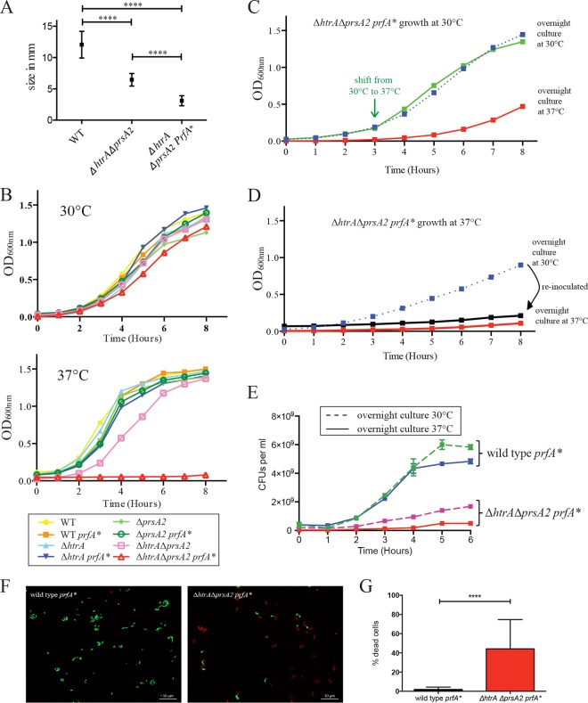
FIG 1.
L. monocytogenes ΔhtrA ΔprsA2::erm prfA* mutants exhibit pronounced growth defects at 37°C. (A) Colony sizes determined on BHI agar after 24 h of growth at 37°C. At least five colonies were measured from two independent experiments. Values that were statistically significantly different (P < 0.0001) by the t test are indicated by a bar and four asterisks. WT, wild type. (B) Bacterial growth as determined by optical density measurement at 600 nm at 30°C and 37°C at the indicated time points. (C and D) Bacterial growth of ΔhtrA ΔprsA2::erm prfA* at 30°C (C) and 37°C (D). (C) Growth at 30°C from an overnight inoculum culture grown at 37°C or 30°C and a temperature shift from 30°C to 37°C at 3 h. (D) Continuous growth at 37°C from an overnight inoculum culture grown at 37°C or 30°C are shown. The latter culture (blue line) was diluted 1:20 and grown again for 8 h at 37°C (black line). (E) Overnight cultures of the wild-type prfA* strain and the ΔhtrA ΔprsA2::erm prfA* mutant were grown at 30°C or 37°C, diluted 1:20, and grown at 37°C for 6 h. Every hour, a sample was taken, serially diluted, and plated for enumeration of CFU per milliliter. Data are representative of the data from two independent experiments. (F) Live/Dead staining of wild-type prfA* and ΔhtrA ΔprsA2::erm prfA* mutants. Overnight cultures were grown at 30°C, diluted 1:20 into fresh medium, and grown for 6 h at 37°C. Micrographs are representative of at least three independent experiments. (G) Enumeration of bacteria from Live/Dead staining. Data summarize results from at least three independent experiments. For each experiment, at least 30 bacteria were counted from 5 to 10 independent fields.

