FIG 6.
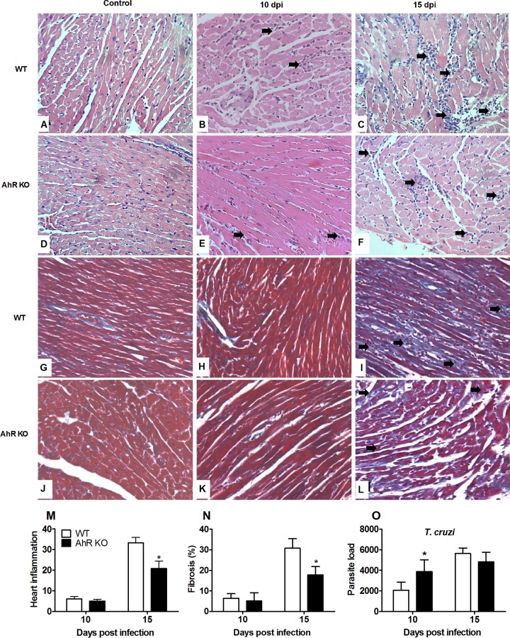
Absence of AhR results in decreased inflammatory cells in heart of T. cruzi-infected mice. WT and AhR KO mice were infected with 1 × 10³ trypomastigotes. Hearts of T. cruzi-infected WT and AhR KO mice were harvested and processed and stained with H&E, and sections were cut from uninfected and T. cruzi-infected WT and AhR-deficient mice to quantify the inflammatory infiltrate and fibrosis. Panels A and D depict the normal histological appearance in uninfected animals. Panels B and E depict discrete and multifocal infiltration of immune cells in myocardium from both infected groups (arrows) at 10 days postinfection (dpi). Panel C depicts multifocal to coalescing and intense inflammatory infiltration in the myocardium (arrows) at 15 dpi. Panel F depicts multifocal and moderate myocarditis (arrows). Panels G and J depict normal myocardium from an uninfected mice. Panels H and K depict minimal deposition of collagen from both infected groups at 10 dpi. (I and L) Intense myocardial fibrosis of an infected WT mouse (I, arrows) and mild fibrosis in AhR KO infected mouse 15 dpi (L, arrows). Original magnification in panels A to F, ×400. Heart inflammation (M), fibrosis (N), and parasite load (O) were scored at 10 and 15 dpi. Data are shown as means ± SEM. *, P < 0.05.

