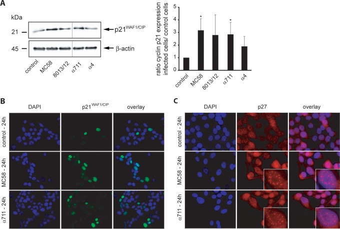
FIG 7.
Pathogenic N. meningitidis strains and carrier isolates increase p21WAF1/CIP1 protein levels in Detroit 562 cells and induce redistribution of p21WAF1/CIP1 and p27CIP1. (A) Cells were either left uninfected (control) or infected with N. meningitidis MC58, 8013/clone12, α711, or α4 (MOI 100) for a 24-h time period and were analyzed for p21WAF1/CIP1 protein expression by Western blot analysis. Band intensities were quantified by densitometric analysis as described for cyclin D1 and cyclin E using ImageJ and normalized to β-actin. The figure shows a representative Western blot with lanes from different areas of the same blot. (B and C) Subcellular distribution of p21WAF1/CIP1 (B) and p27CIP1 (C) in N. meningitidis-infected Detroit 562 cells assessed by immunofluorescence microscopy. Colors in panel B: green, p21WAF1/CIP1 protein stained with specific antibody and secondary antibody conjugated with Alexa Fluor 488; blue, nuclei stained with DAPI; light green, colocalization. Colors in panel C: red, p27CIP1 stained with specific antibody and secondary antibody conjugated with Cy3; blue, nuclei stained with DAPI.

