Figure 1.

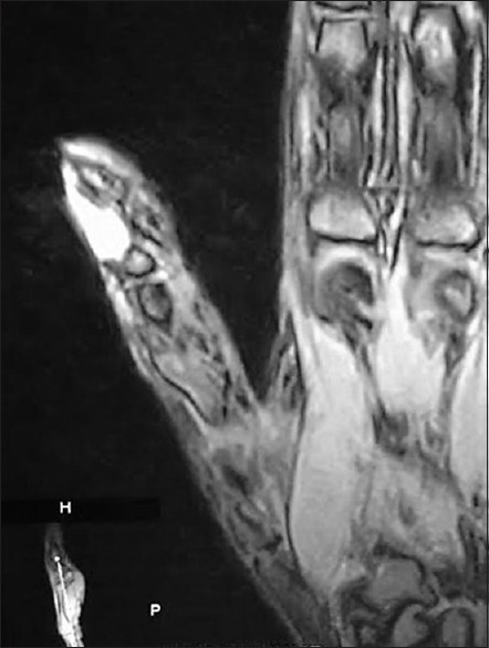
Magnetic resonance imaging showing a tumor at the subungual aspect of distal phalanx which is hyperintense on T2 weighted images

Magnetic resonance imaging showing a tumor at the subungual aspect of distal phalanx which is hyperintense on T2 weighted images