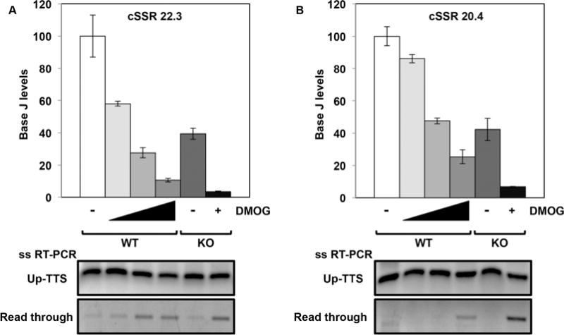
Fig. 3.

Levels of J remaining in the H3.V KO are sufficient for terminating transcription.
A and B. The levels of J at the TTS of two representative cSSRs were determined by J IP-qPCR analysis in WT cells with increasing DMOG concentrations (0, 1, 2.5 and 5mM) and in H3.V KO cells in the absence and presence of 5 mM DMOG. At cSSR 22.3, reduction of J is statistically significant in each condition relative to WT. At cSSR 20.4, reduction of J is statistically significant in each condition relative to WT except WT+1mM DMOG. All statistical significance was determined using two-tailed Student’s t-test and a P-value <0.05. Below, strand-specific RT-PCR analysis of each of the cSSRs as described in Fig. 2B.
