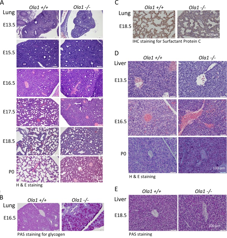
FIG 2.
Phenotypic analysis of Ola1+/+ and Ola1−/− embryos. (A) H&E staining of Ola1+/+ and Ola1−/− lungs from E13.5 to E18.5 embryos and pups on the day of birth (P0). (B) Periodic acid-Schiff (PAS) staining of lungs sections from E16.5 embryos. (C) Immunohistochemical staining with the antibody against SP-C in lung sections from E18.5 embryos. The nuclei were counterstained using eosin. (D) H&E-stained histological sections of livers obtained from Ola1+/+ and Ola1−/− embryos at E13.5 and E16.5 and from neonates (P0). (E) PAS staining of liver sections of embryos obtained at E16.5.

