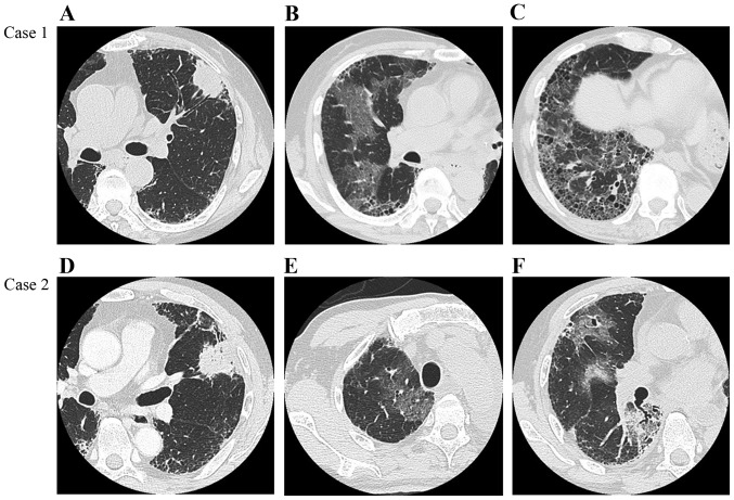
Figure 1.
Chest high-resolution computed tomography (HRCT) images. Case 1: (A) Chest HRCT scan prior to surgery showing a lobulated subpleural mass in the left upper lobe, measuring 4 cm in diameter, on a background of reticulation and honeycombing, predominantly in the bilateral lower lobes. (B and C) Chest HRCT scans on day 5 after surgery showing multifocal ground-glass opacities (GGO), predominantly in the non-operated right lung. Case 2: (D) Chest HRCT scan prior to surgery showing a subpleural mass in the left upper lobe exhibiting spiculation and air bronchogram, measuring 7.5 cm in diameter, with reticulation and faint honeycombing, predominantly in the bilateral lower lobes (usual interstitial pneumonia pattern). (E and F) Chest HRCT scans on day 10 after surgery showing multifocal GGO predominantly in the non-operated right middle and lower lobes.

