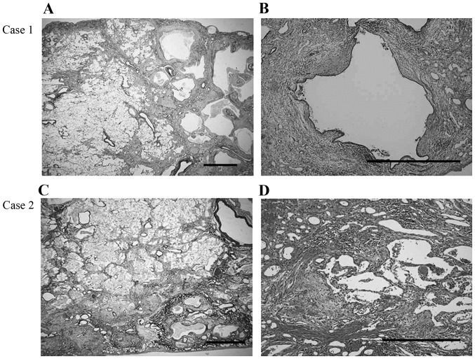
Figure 2.
Microscopic appearance of the resected lungs. Case 1: (A) Heterogeneous interstitial fibrosis with honeycombing with a subpleural and perilobular distribution pattern, alternating with areas of normal lung tissue (Elastic van Gieson stain; scale bar, 1 mm). (B) Fibroblastic foci are sporadically present in a dense collagen fibrosis background and infiltration by abundant lymphocytes and neutrophils is observed (hematoxylin and eosin stain; scale bar, 1 mm). Case 2: (C) Background pattern of usual interstitial pneumonia with fibrotic changes, predominantly distributed in the subpleural and perilobular areas, and an abrupt transition between almost-normal alveolar septa and dense fibrosis with architectural disruption (Elastic van Gieson stain; scale bar, 500 mm). (D) Prominent fibroblastic foci and infiltration by lymphocytes and neutrophils in the interstitium (hematoxylin and eosin stain; scale bar, 500 mm).

