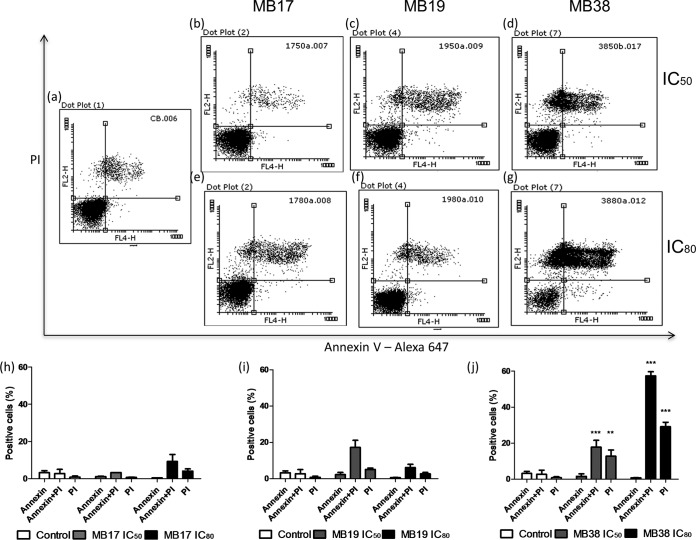
FIG 3.
Analysis of extracellular exposure of phosphatidylserine by annexin V/propidium iodide labeling. The parasites were treated with MB17, MB19, and MB38 at concentrations corresponding to the IC50s (0.5 ± 0.13, 3.7 ± 0.29, and 8.3 ± 1.49 μM, respectively) and IC80s (1.5 ± 0.5, 8.0 ± 0.94, and 16 ± 3.2 μM, respectively) for 5 days. After this period, the parasites were labeled with annexin V and propidium iodide and analyzed by flow cytometry as follows: untreated epimastigotes (a), parasites treated with MB17 at concentrations corresponding to the IC50 (b) and IC80 (e), parasites treated with MB19 at concentrations corresponding to the IC50 (c) and IC80, (f), and parasites treated with MB38 at concentrations corresponding to the IC50 (d) and IC80 (g). Quantitative analysis of the parasites treated with MB17, MB19, and MB38, as indicated, are also shown. One-way ANOVA followed by a Tukey posttest was used for statistical analysis to compare the treatments to values in the respective control. ***, P < 0.001; **, P < 0.01 (Tukey posttest).

