FIG 3.
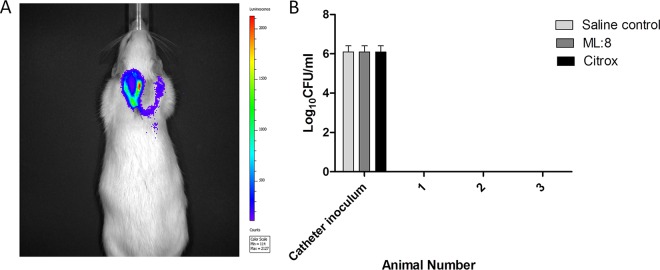
Monitoring infection development and dissemination. Optimized doses of high-inoculum (106 CFU/ml) MRSA USA300 lux in 40 μl were injected into catheters, and infection was allowed to develop for 5 days. (A) An IVIS imaging system was used to monitor the development of infection. Bioluminescence activity in a Sprague-Dawley rat was acquired using an IVIS 100 camera at 5 days postinfection. The color scale indicates the degree of luminescence, from high (red) to low (purple). A representative animal is shown (n = 3). (B) Catheter inoculation and dissemination into blood were assessed at the experiment endpoint. Animals were euthanized at day 9, blood was harvested, and log10 CFU counts were determined. Assays were performed in triplicate, and the data represent mean log10 CFU per milliliter and SD.

