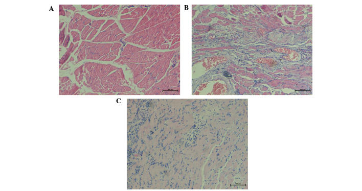
Figure 2.
Hematoxylin-eosin staining of representative tissue samples collected at 4 weeks after surgery in the (A) control, (B) osteomyelitis model and (C) decorin-treated groups (magnification, ×100). The control group showed well-arranged muscle fibers and few microvessels. In the osteomyelitis group, tissue was characterized by evident hyperplasia, with an increased number of microvessels, chronic inflammation, and irregularly-arranged nodular, circular or whorled muscle fibers. The decorin-treated group showed a gradual reduction in blood vessels and chronic inflammation, with well-arranged muscle fibers.

