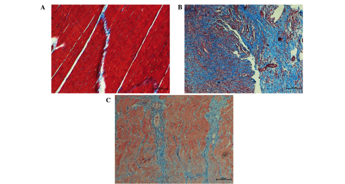
Figure 3.
Masson's trichrome staining representative tissue samples in different groups (magnification, ×200). (A) In the control group, red muscle fibers were regularly arranged with presence of few detached collagen deposits. (B) In the osteomyelitis group, atrophic muscle fibers were irregularly arranged and increased blue collagen areas was present. (C) In the decorin-treated group, muscle fibers were relatively regular and blue areas of collagen deposition were reduced.

