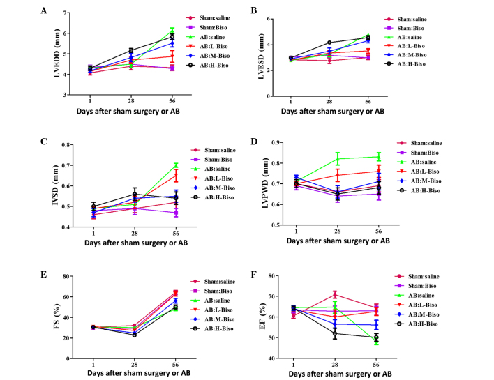
Figure 1.
Echocardiographic parameters at different time-points. (A) Left ventricular end-diastolic diameter (LVEDD), (B) left ventricular end-systolic diameter (LVESD), (C) left ventricular septum diastolic (IVSD), (D) left ventricular posterior wall diameter (LVPWD), (E) fractional shortening (FS) and (F) ejection fraction (EF). Data are presented as mean ± standard error of the mean (n=12–16 in each group) and were analyzed with 2-way repeated-measures analysis of variance. Bonferroni correction was applied for multiple comparisons. (A-F) P<0.01 for H-Biso and M-Biso vs. the other four groups at 8 weeks (56 days), but no significant difference between the M-Biso and H-Biso groups. AB, aortic banding; Biso, bisoprolol; L, low-dose; M, middle-dose; H, high-dose.

