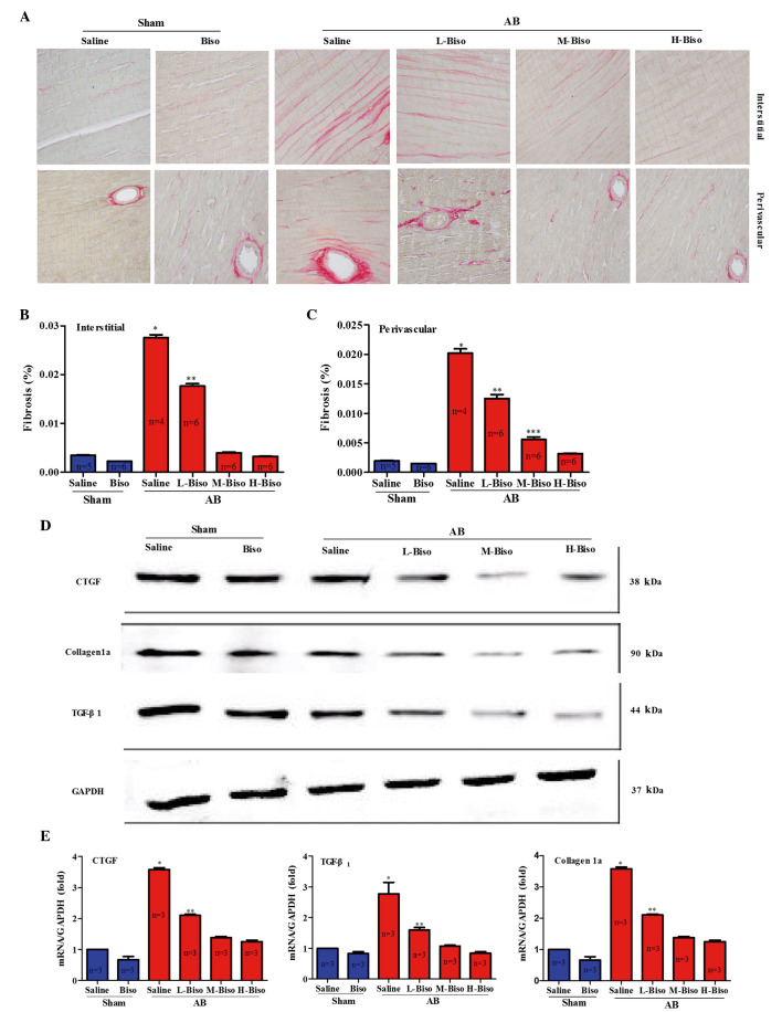
Figure 4.
Bisoprolol attenuates cardiac fibrosis in response to pressure overload in vivo. (A) Picrosirius red staining of interstitial (top) and perivascular (bottom) regions of left ventricles of C57BL/6J mice following treatment of saline or bisoprolol at 8 weeks after sham or aortic banding (AB) surgery. Quantification of collagen volume fraction from (B) interstitial and (C) perivascular regions (n=4–6 per group). (D) Representative western blots of connective tissue growth factor (CTGF), tissue growth factor (TGF)-β1 and collagen 1a at 8 weeks after sham or AB surgery. (E) Quantitative mRNA expression levels of CTGF, TGF-β1 and collagen 1a in the groups (n=3 per group). Values represent the mean ± standard error of the mean. *P<0.05 vs. all other groups. **P<0.05 vs. M-Biso and H-Biso AB groups; ***P<0.05 vs. H-Biso AB groups. Biso, bisoprolol; L, low-dose; M, middle-dose; H, high-dose; GAPDH, glyceraldehyde 3-phosphate dehydrogenase.

