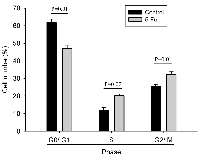
Figure 1.
Cell cycle analysis of PLC/RAF/5 and PLC/RAF/5/5-Fu cells. Quantification of cell proliferation using propidium iodide staining by flow cytometry (n=3). Data were expressed as the percentage of cells in each phase. Control, PLC/RAF/5 cells; LC/RAF/5/5-Fu cells, 5-Fu-treated cells. 5-Fu, 5-fluorouracil.

