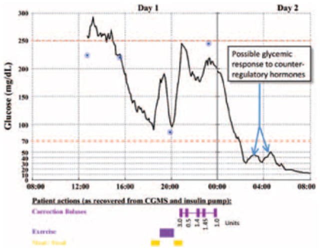
Figure 4.

Glucose levels captured by the retrospective CGM for the evening before and the morning of the patient’s death. The calibrations measured and entered by the patient are represented by the 4 circles. The timings of the patient’s meals, exercise, and correction insulin boluses are represented by the bars along the bottom of the graph. The precipitous decrease in glucose level after the correction doses can be observed to start just after midnight, and possible counterregulatory efforts are noted once the glucose level declined to below 30 mg/dL shortly after 2 am.
