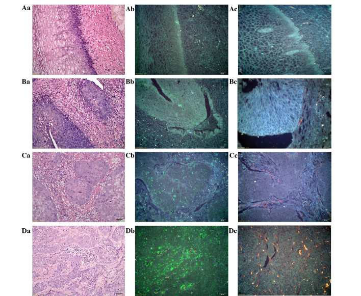
Figure 2.
Dynamic changes in the tumor microenvironment during CC progression. (Aa) H&E staining, (Ab) TAM staining with QDs-525 and (Ac) tumor neo-vessel staining with QD-585, in normal tissue. (Ba) H&E staining, (Bb) TAM staining with QDs-525 and (Bc) tumor neo-vessel staining with QD-585, in CC in situ tissue. (Ca) H&E staining, (Cb) TAM staining with QD-525 and (Cc) tumor neo-vessel staining with QD-585, in well-differentiated CC. (Da) H&E staining, (Db) TAM staining with QD-525 and (Dc) tumor neo-vessel staining with QD-585, in poorly-differentiated CC. Aa-Ac, Ba and Bb, Ca-Cc and Da-Dc: Magnification, ×200; and Bc: Magnfication, ×400. TAMs, tumor-associated macrophages; QD, quantum dot; CC, cervical cancer; H&E, hematoxylin and eosin.

