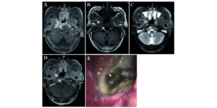
Figure 2.
MRI examination performed on a 64-year-old man diagnosed with NPC with T3N3M0, who underwent IMRT with two courses of neo-adjuvant chemotherapy and received a partial response of short-term therapeutic effects of chemotherapy. (A) Transverse contrast-enhanced T1-weighted MRI revealed enhanced tumour tissue invading the musculus longus capitis, parapharyngeal space and medial pterygoid muscle on the right side of the nasopharyngeal cavity (as indicated by the arrows). (B) Transverse T1-weighted MRI revealed a low-signal area in the initial tumour bed at 1 month following the completion of RT (demonstrated by the arrows). (C) Transverse T2-weighted MRI revealed a mixed signal area in the initial tumour bed (shown by the arrows). (D) Transverse contrast-enhanced, T1-weighted MRI revealed a non-enhanced area, which showed the ulcer invading, but not exceeding, the muscle tissue (demonstrated by the arrows). The ulcer was categorised as a moderate-grade UPRNN. (E) The ulcer was located on the right side of the nasopharyngeal cavity (indicated by the arrow). RT, radiotherapy; MRI, magnetic resonance imaging; NPC, nasopharyngeal carcinoma; IMRT, intensity-modulated RT; UPRNN, ulcer of post-radiation nasopharyngeal necrosis.

