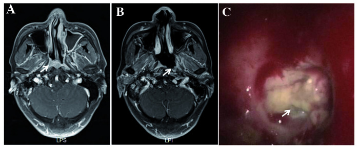
Figure 4.
MRI examination performed on a 69-year-old man diagnosed with NPC with T3N2M0/rT3N0M0, who underwent RT twice. The second RT was IMRT with two courses of neo-adjuvant chemotherapy. The ulcer occurred at 4 months following the completion of IMRT. The patient was diagnosed with RIMU, not UPRNN. (A) Transverse contrast-enhanced, T1-weighted MRI revealed enhanced tumour tissue invading the parapharyngeal space and medial pterygoid muscle on the left side of the nasopharyngeal cavity (shown by the arrow). (B) Transverse contrast-enhanced, T1-weighted MRI revealed a large ulcer with non-enhancement on the right side of the nasopharyngeal cavity at 4 months following the completion of RT, which was not previously occupied by clear tumour tissue (shown by the arrow). (C) The ulcer was located on the left side of the nasopharyngeal cavity (indicated by the arrow). RT, radiotherapy; MRI, magnetic resonance imaging; NPC, nasopharyngeal carcinoma; IMRT, intensity-modulated RT; RIMU, mucosal ulcer induced by radiation; UPRNN, ulcer of post-radiation nasopharyngeal necrosis.

