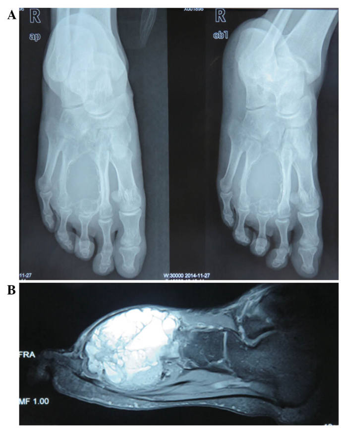
Figure 1.

(A) Radiograph showing a lytic lesion in the entire third metatarsal, leaving the cortex as a thin shell with a ‘finger in a balloon’ appearance. (B) T2-weighted magnetic resonance imaging sequence showing deformity of the involved metatarsal bone with fluid-fluid levels.
