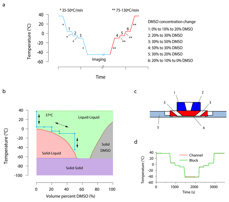
Figure 1. Protocol for reversible cryo-arrest on a microscope stage.
(a) Time course of the cryo-arrest protocol. Temperature is changed stepwise between the physiological temperature of 37°C and the fixation temperature of -45°C and DMSO concentration is adapted as indicated on the right side of the figure (1-6). Media was exchanged with 3 μL s-1 to avoid cell detachment. * and ** represent the cooling and warming speeds indicated in the figure. (b) Binary phase diagram of DMSO-water mixtures (data adapted from 8) including the course of the cryo-arrest protocol (blue line, arrows). The green area indicates the temperature range in which the mixture is a stable liquid (and solidification is thermodynamically forbidden) (c) Side view sketch of the central part of the cryo-stage. (1) silver block with temperature control; (2) aluminum block; (3) medium inlet and outlet; (4) 100 μm thick channel with sample on a cover slide; (5) insulating polyvinyl chloride plate adapted for commercial microscopy stages. (d) Temperature recording during the course of an experiment. The temperature was measured directly in the channel with the help of a 50 μm thick thermocouple (red line). This temperature was compared to the temperature read out at the aluminum block of the stage (green line), as routinely performed during every experiment.

