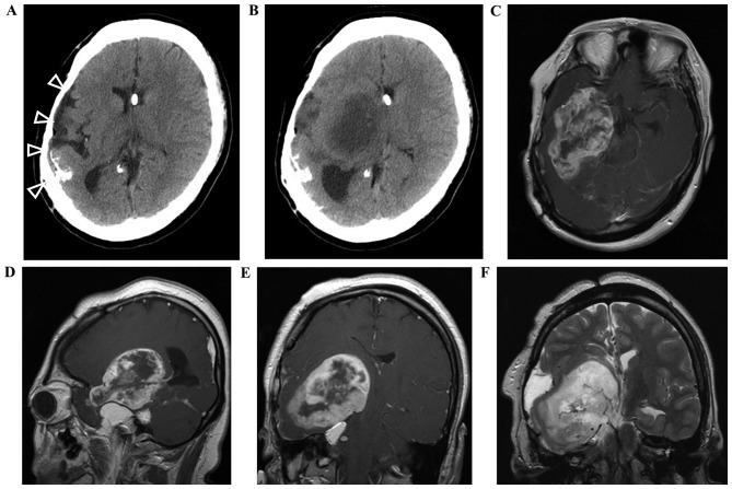
Figure 2.
Computed tomography (CT) and magnetic resonance imaging (MRI) scans of the tumor. (A) No tumor was observed on a CT scan performed ~1 year prior to onset. Atrophy and calcification (arrowheads) in the right hemisphere, which are a common characteristic of encephalocraniocutaneous lipomatosis, are visible. (B) A low-density mass was observed in the right basal ganglia on a CT scan performed at the time of onset. (C-E) Gadolinium-enhanced MRI revealed a ring-enhanced tumor extending from the right temporal lobe to the basal ganglia adjacent to the intracranial lipoma. (F) A small amount of perifocal edema was present in the vicinity of the tumor.

