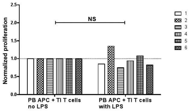
Figure 4.

Proliferation of TI T cells when incubated with PB APCs. Six patients were included and each bar represents one subject. Purified blood APCs were pre-incubated without or with 2 µg/ml LPS for 3 days and then added to autologous tumor leukocyte culture. The cells were then labeled with CFSE and stimulated with anti-CD3 antibodies for 6 days. Data were normalized against the results without LPS in each patient (paired t-test). NS, not significant; TI, tumor-infiltrating; PB, peripheral blood; APC, antigen-presenting cell; LPS, lipopolysaccharide.
