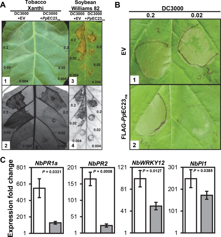
Fig 1. PpEC23 suppresses HR induced by Pst DC3000.
A. HR on leaves of N. tabacum cv. Xanthi (A1, A2) and G. max cv. Williams 82 (A3, A4) infiltrated with Pst DC3000 carrying empty pEDV6 vector (EV) (left half) and Pst DC3000 carrying pEDV6::PpEC23ns (right half). Leaves were infiltrated with bacteria at OD 600 nm = 0.2, 0.02, and 0.004 (approximately equal to 108, 107 and 2x10⁶ CFU (colony forming units) / mL, respectively [32]). Leaves were photographed at 18 h post infiltration (hpi) for tobacco (A1) and at 30 hpi for soybean (A3) and then stained with trypan blue to detect non-viable cells (A2, A4). B. HR on leaves of N. benthamiana stably transformed with EV (B1) and FLAG-PpEC23ns (B2) infiltrated with Pst DC3000. Leaves were infiltrated with bacteria at OD 600 nm = 0.2 (left), 0.02 (right). Leaves were photographed at 18 hpi. Representative images are shown (n ≥ 8). C. The expression fold change of immune marker genes PR1a, PR2, WRKY12, and PI1 in the leaves of N. benthamiana stably transformed with EV (white bars) and FLAG-PpEC23ns (gray bars) at 16 hpi with Pst DC3000. NbAct1 was used as the internal reference gene. A t-test was performed for each pair-wise comparison and the P value for each comparison is shown. Four biological and four technical replicates were performed.

