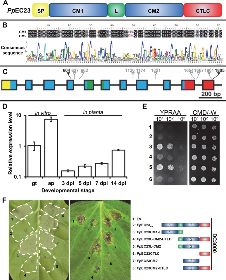
Fig 2. PpEC23 is a modular SSCRP that is present in diverse P. pachyrhizi isolates.
A. Diagram depicting the different motifs of PpEC23. SP, signal peptide; CM1, ten-cysteine motif 1; L, linker; CM2, ten-cysteine motif 2; CTLC, C-terminal low complexity motif. B. Sequence alignment of the two CMs in PpEC23 (top) and the consensus sequence of CM for all members of cluster 112, generated by WebLogo (bottom). C. Genomic structure of the PpEC23 gene. Colors of the exons shown as blocks correspond with motif colors in panel A. Arrows show the SNPs, together with the nucleotide position, among ten geographically distinct isolates. Hollow gray, solid gray and black arrows show the intron SNPs, synonymous SNPs, and nonsynonymous SNPs, respectively. D. Transcript levels of PpEC23 at different developmental stages. Transcript levels were normalized to the expression of the fungal reference genes, RPS14 and PDK. Three independent biological and four technical replicates were performed. Error bars = standard deviation. E. The PpEC23 signal peptide directs protein secretion in yeast. Fusion constructs of PpEC23 in the secretion signal trap plasmid: 1) pSuc2t7M13ori::PpEC23, 2) pSuc2t7M13ori::PpEC23ns, 3) pSuc2t7M13ori::PpEC23Sp-CM1-L, 4) pSuc2t7M13ori::PpEC23L-CM2-CTLC, 5) pSuc2t7M13ori::PpEC23Sp, 6) pSuc2t7M13ori EV control. YPRAA and CMD/-W indicates the selective and non-selective medium, respectively. Numbers below indicate the fold dilution of the yeast strains. F. Symptoms on leaves of N. tabacum cv. Xanthi (left) and G. max cv. Williams 82 (middle) infiltrated with Pst DC3000 carrying EV, PpEC23ns, and various truncated constructs of PpEC23. The name and structural diagram of each construct is provided in the right panel. Leaves were infiltrated with bacteria at OD 600 nm = 0.02. Leaves were photographed at 18 hpi for tobacco (left) and at 30 hpi for Williams 82 soybean (middle). Representative images are shown (n ≥ 8).

