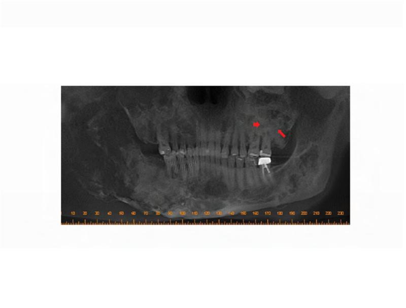
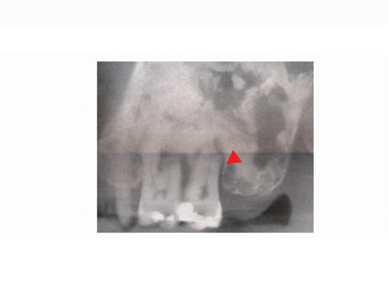
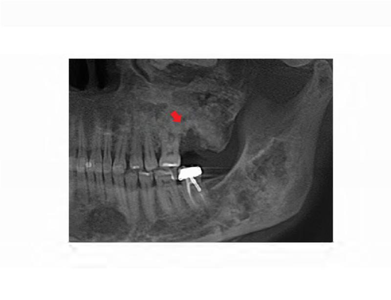
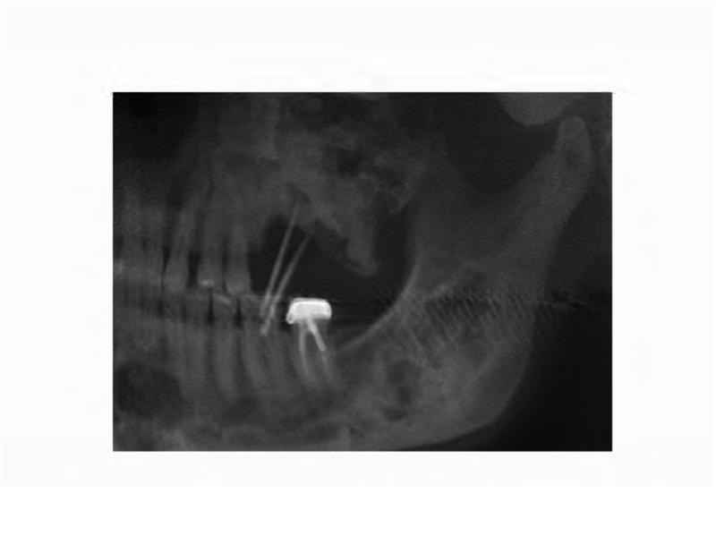
Figure 2. Case 1 dental images.
A. Pre-operative panoramic radiograph taken prior to extraction of the left maxillary second molar and three months prior to initial presentation of osteonecrosis. Both the left maxillary first and second molars are located within fibrous dysplasia bone (red arrows). B. Periapical radiograph taken prior to extraction of the left maxillary second molar (ADA #15). Note the perio-endo lesion associated with the right maxillary second molar (red arrowhead). C. A panoramic radiograph taken four months post-extraction of the left maxillary second molar shows bone resorption and osteosclerosis of the lesion (red arrow). D. Gutta percha points within the persistent fistula.


