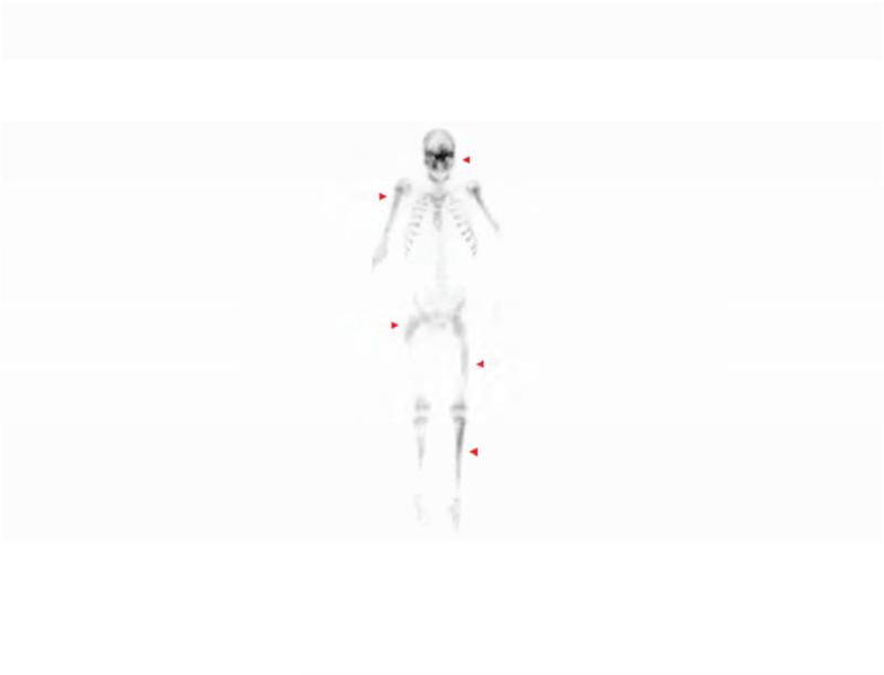
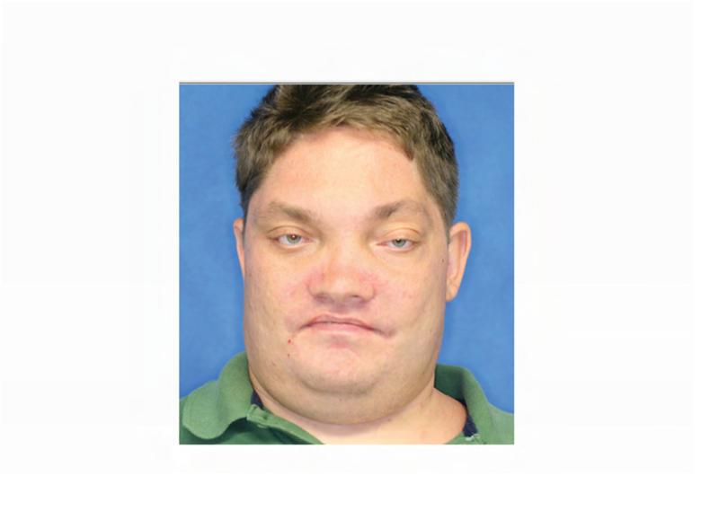
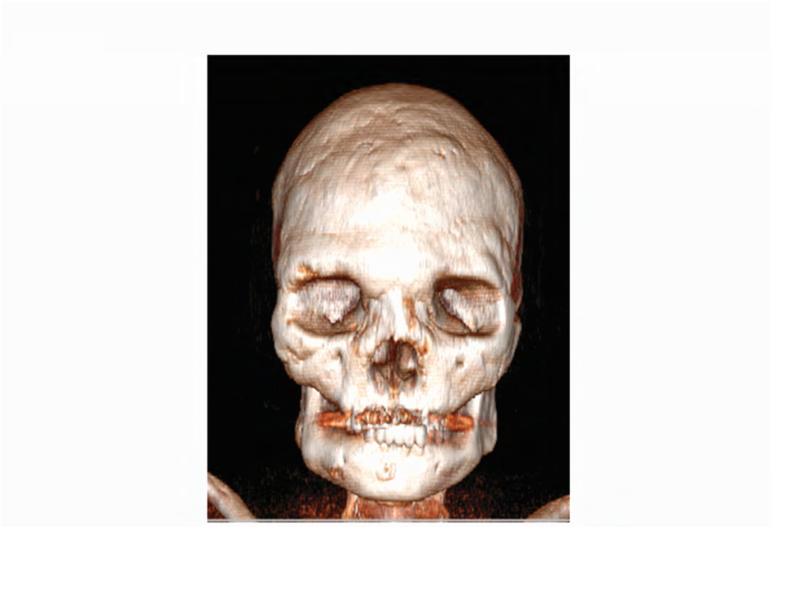
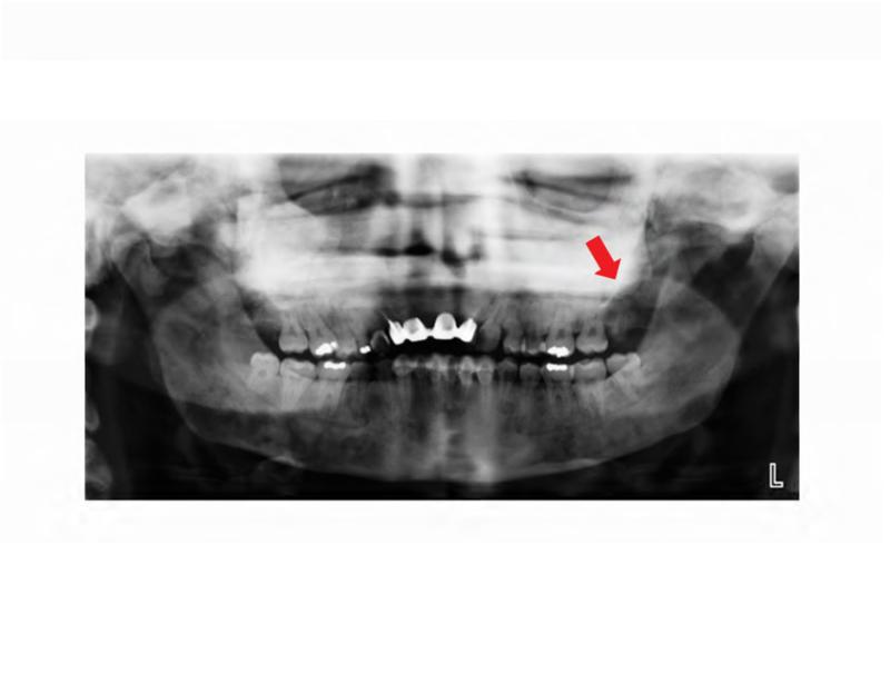
Figure 3. Case 2 clinical and dental images.
A. Technetium-99 bone scan highlighting areas of increased radiotracer uptake in bone (red arrowheads) affected by fibrous dysplasia in the skull and bilateral femora, humeri, and fibulae. B. Clinical photo depicting facial dysmorphism resulting from mild vertical dystopia, and diffuse expansion of maxillary and mandibular fibrous dysplasia. C. Three-dimensional computed tomography reconstruction reveals diffuse craniofacial involvement with fibrous dysplasia. D. Panoramic radiograph demonstrating the region of significantly decayed left maxillary third molar (red arrow).


