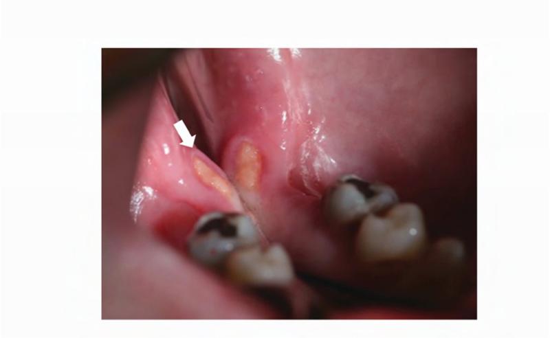
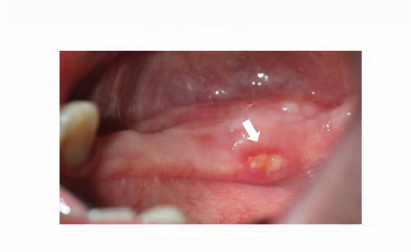
Figure 6. Case 3 intraoral photographs.
Photographs were taken 2 weeks after initial ONJ presentation. A. Right posterior mandible with exposed bone along the lingual alveolar ridge (image is reflected in mirror). B. Patient's left mandibular lingual alveolus, reflected in mirror. Area of exposed bone is present along the posterior mylohyoid ridge.


