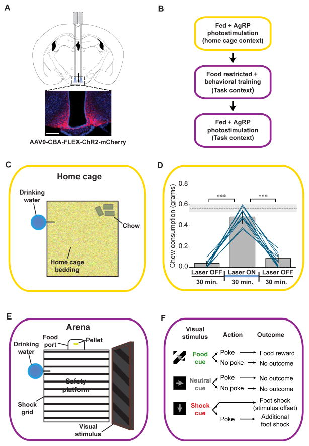
Figure 1. Experimental design for AgRP neuron stimulation in freely moving mice in the home cage and during a visual discrimination task.
(A) Selective expression of ChR2-mCherry in AgRP neurons in the arcuate nucleus of the hypothalamus. Optic fibers were implanted either uni- or bilaterally (see Supplemental Experimental Procedures and Figure S2C). Scale bar, 200 μm. (B) Flow chart illustrating order of experiments. Once photostimulation-evoked feeding in the home cage was confirmed in fed mice, mice were food-restricted, trained on a visual discrimination task, returned to ad libitum access to food, and then tested for photostimulation-evoked discrimination behavior. (C) Schematic showing the home cage setting used to test for AgRP stimulation-evoked feeding. (D) Food intake before, during, and after unilateral AgRP neuron photostimulation in the home cage setting. Lines represent mean intake for all individual mice used in all subsequent photostimulation experiments (n = 11); bars represent grand average ± SE. Paired t-tests, ***p < 0.001; dashed line represents mean ± SE chow consumption in mice fasted for 24 hours. (E) Schematic showing the behavioral arena used for visual discrimination training. (F) Schematic of visual discrimination task stimulus-response contingencies. See also Figure S1 and Movie S1.

