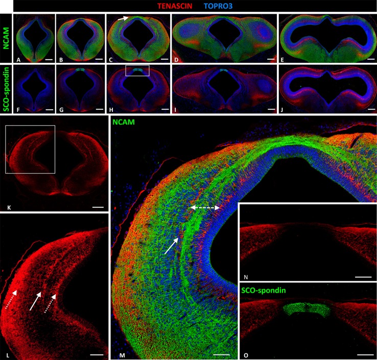
Figure 2.
Expression of tenascin, SCO-spondin, and NCAM in frontal sections of HH29 chick diencephalon and mesencephalon. (A–E) Confocal images showing the co-localization of NCAM (green) and tenascin (red) in the region between prosomere 2 and the mesencephalon. The PC is located in the medial region of prosomere 1 (arrow in C). (F–J) Confocal images showing co-localization of SCO-spondin (green) and tenascin (red) in frontal sections between prosomere 2 and the mesencephalon, showing the presence of SCO-spondin only in the roof plate of prosomere 1 (G–I). (K–M) Higher magnification of (C) showing tenascin arrangement into three columns (arrows in L). The axonal fascicles of the PC extended inside a corridor (dotted double-arrow in M) delimited by the tenascin columns. (N,O) Higher magnification of the area framed in (H) showing the absence of tenascin in the roof plate, coinciding with the expression of SCO-spondin. TOPRO 3 (blue) was used as nuclear counterstain. Scale bars = 200 μm in (A–K); 100 μm in (L); and 50 μm in (M–O).

