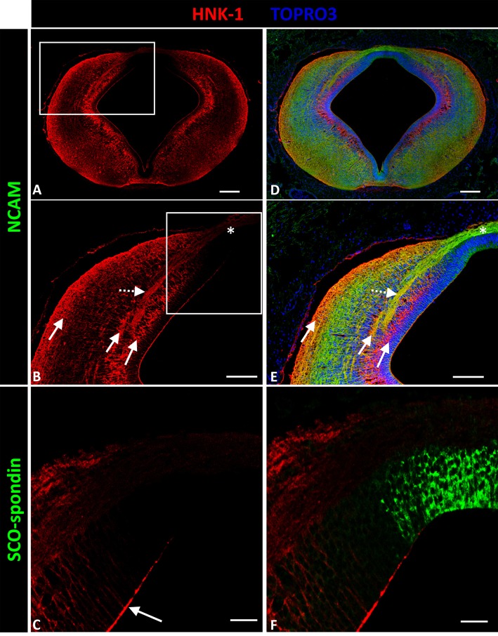
Figure 3.
Expression of HNK-1, SCO-spondin, and NCAM in frontal sections of chicken prosomere 1 at stage HH29. (A,B,D,E) Confocal images showing co-localization of NCAM (green) and HNK-1(red). HNK-1 is arranged into three columns (solid arrows in B). The axonal fascicles of the PC extended inside a corridor formed by these columns. The axons of the PC are immunopositive for HNK-1 in the lateral regions (dotted arrow in E), but the immunoreactivity highly diminishes in the dorsal region and in the roof plate (asterisk in B,E). (C,F) Confocal images showing co-localization of SCO-spondin (green) and NHK-1(red) in an area equivalent to the area framed in (B), showing the absence of HNK-1 in the roof plate, coinciding with the expression of SCO-spondin. HNK-1 is present in the apical region of the neuroepithelial cells (arrow in C), with the exception of roof plate cells positive for SCO-spondin. TOPRO 3 (blue) was used as nuclear counterstain. Scale bars = 100 μm in (A,B,D,E); 25 μm in (C,F).

