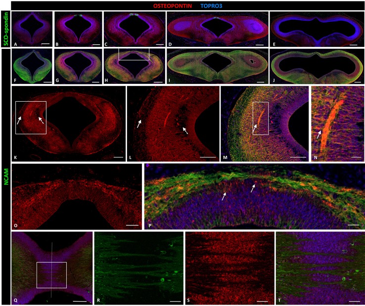
Figure 4.
Expression of osteopontin, SCO-spondin, and NCAM in frontal and horizontal sections of chicken diencephalon and mesencephalon at stage HH29. Double immunohistochemistry with anti-SCO-spondin (A–E, green) or NCAM (F–J, green) and osteopontin (red) in the region between prosomere 2 and mesencephalon. Osteopontin was highly expressed in prosomere 1 (B–D) but not in prosomere 2 (A) or the mesencephalon (E). (K) Expression of osteopontin in the middle region of prosomere 1. (L–N) Magnification of the region framed in (K) showing the wide expression of osteopontin, with the exception of the periventricular and superficial strata (dotted and continuous arrows, respectively, in K,L). (N) Higher magnification of the area framed in (M) showing the co-localization of NCAM and osteopontin in the axonal fascicle. (O,P) Magnification of the area framed in (H) showing osteopontin expression in the dorsal region and in the roof plate, between the PC axons. (Q–T) Horizontal sections of the roof plate of prosomere 1 showing the axons crossing the midline (dotted line) and immersed in a osteopontin-rich medium. (R–T) magnification of the area framed in (Q). TOPRO 3 (blue) was used as nuclear counterstain. Scale bars = 200 μm in (A–J); 100 μm in (K); 50 μm in (L,M,O,Q); 10 μm in (N,P,R,S).

