Figure 5.
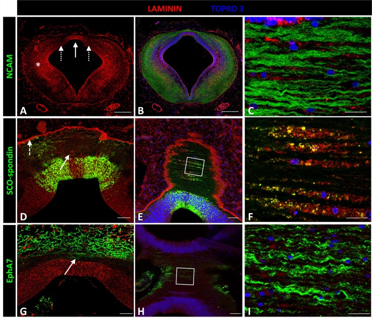
Expression of laminin, SCO-spondin, EphA7, and NCAM in frontal and horizontal sections of chicken prosomere 1 at stage HH29. (A–C) Double immunohistochemistry with anti-NCAM (green) and laminin (red) showing the broad expression of this ECM molecule, especially in the lateral region (asterisk in A) and in the roof plate (solid arrow in A). (C) Coronal section of the roof plate reveals the presence of laminin in the ECM surrounding the axons. (D) Frontal section of the roof plate showing the presence of laminin in the midline (solid arrow) and in the external limiting membrane (dotted arrow). (E,F) Horizontal images of the prosomere 1 roof plate showing that laminin colocalizes with SCO-spondin in the ECM surrounding the axons. (F) Magnification of the area framed in (E). (G–I) Double immunohistochemistry with anti-EphA7 (green) and laminin (red) showing the expression of EphA7 in the dorsal midline cells (arrow in G). (H,I) Horizontal images of the prosomere 1 roof plate showing that laminin is in close contact with the basal prolongations of the midline cells positives for EphA7. (I) Magnification of the area framed in (H). TOPRO 3 (blue) was used as nuclear counterstain. Scale bars = 200 μm in (A,B); 20 μm in (D,E); 10 μm in (C,F).

