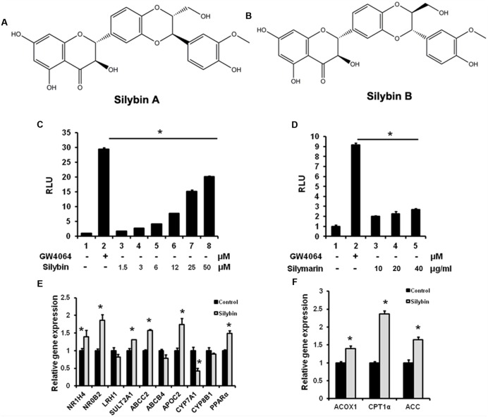
FIGURE 1.
Silybin and silymarin activate FXR transactivity in vitro. (A,B) Structure of Silybin A and Silybin B. Silybin (C) and silymarin (D) activate FXR transactivity. HEK293T cells were co-transfected with phFXR, phRXR expression plasmids, and FXR-dependent reporter (EcRE-LUC) for 24 h and treated with the FXR agonist GW4064 (10 μM), control (DMSO), silybin (1.5, 3, 6, 12, 25, 50 μM) and silymarin (10, 20, and 40 μg/ml) for another 24 h. The relative luciferase activities (RLU) were measured by comparison to renilla luciferase activities. (E,F) The relative gene expression levels in silybin-treated (25 μM) and control (DMSO) HepG2 cells. Beta-ACTIN was used as an internal control for normalizing the mRNA levels. The results represent three independent experiments, and data are presented as means ± SEM. (n = 3). ∗P < 0.05 vs. HEK293T or HepG2 control.

