
Figure 5.
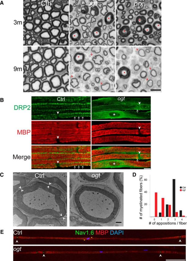
Abaxonal apposition formation and internodal length are abnormal in OGT-SCKO nerves. A, Representative toluidine blue-stained semithin cross sections of sciatic nerves from Ctrl, OGT-SCKO (ogt), and Prx-null (prx) mice at 3 and 9 months of age. Note the decrease in the number of axons in the mutant animals and the presence of tomacula (asterisks) and thinly myelinated fibers (arrows). Scale bar, 10 μm. B, Representative confocal microscopic images of teased sciatic nerve fibers from 1-month-old OGT-SCKO and control mice labeled for DRP2 (green) and MBP (red). Abaxonal appositions (arrows). Note the diffuse DRP2 staining and paucity of appositions in OGT-SCKO nerve. The nodes of Ranvier (arrowheads) and the SC nucleus (asterisk) are labeled. Scale bar, 10 μm. C, Electron micrograph showing myelinated fibers of sciatic nerve in a cross section from 1-month-old control and OGT-SCKO mice. Pairs of arrowheads mark the abaxonal appositions residing between the compact myelin and SC plasma membrane. Note the lack of abaxonal appositions in the OGT-SCKO fiber. Scale bar, 1 μm. D, Quantification of abaxonal appositions displayed as average per myelinated fiber in nerves from B. Note the dramatic decrease in appositions in OGT-SCKO mice. Approximately 30 fibers per mouse were analyzed; n = 3 mice per genotype. E, Representative confocal microscopic images depicting the entire internodal length of myelinated fibers from 3-month-old control and OGT-SCKO nerves labeled for NAV1.6 (green), MBP (red), and DAPI (blue). Note the decrease in intermodal length in the OGT-SCKO nerve (n = 3 mice per genotype, 10 fibers per mouse). Arrowheads mark the node of Ranvier. Scale bar, 100 μm.