
Figure 6.
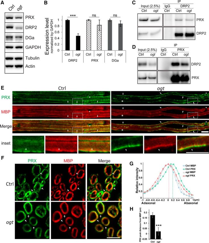
O-GlcNAcylation is required for proper PRX localization but does not affect DRP2–PRX interaction. A, Western blot analysis of PRX, DRP2, and α-Dystroglycan in sciatic nerves from 1-month-old control and OGT-SCKO mice. DGa, α-Dystroglycan; GAPDH, glyceraldehyde-3-phosphodehydrogenase; Actin, β-actin; Tubulin, β-tubulin. Note the decrease in the DRP2 level in the OGT-SCKO nerve. B, Quantification of PRX, DRP2, and DGa in A. Normalized by GAPDH (n = 3 mice per genotype). Data are represented as mean ± SEM. Student's t test, ***p < 0.001); ns, not significant. C, D, Coimmunoprecipitation analysis of interaction between PRX and DRP2. Nerve lysates were immunoprecipitated for DRP2 (C) or PRX (D). The bound partner was detected by Western blotting with indicated antibody. IP, Immunoprecipitation; IgG, preimmune rabbit sera. A total of 2.5% of the lysate was used for input control (Input). Note that the PRX–DRP2 interaction was maintained in OGT-SCKO nerve lysates. E–H, Localization of PRX in Ctrl and OGT-SCKO (ogt) sciatic nerves. E, F, Representative confocal microscopic images of teased fibers (E) and cross sections (F), stained for PRX (green) and MBP (red). E, In teased fiber preparations, PRX distributes as elongated coarse streaks (arrows) along the myelin sheath in OGT-SCKO nerves, whereas it localizes to apposition-like features (arrows) in controls. The nodes of Ranvier (arrowheads) and the SC nucleus (asterisk) are labeled. The area enclosed by the dashed line (inset) was enlarged to highlight appositions in control and the corresponding area in the mutant. F, In cross sections, PRX concentrates peripherally to MBP (arrows) in control nerves, whereas PRX and MBP overlap extensively in OGT-SCKO nerves. G, Line-plot profile of PRX and MBP distribution across the myelin sheath in F. Pixel intensities of PRX and MBP on a straight line drawn across the myelin sheath were first aligned according to the peak coordinate of MBP per genotype, then averaged and normalized to the max intensity per protein. Note that PRX and MBP extensively overlap in OGT-SCKO nerves but are segregated in control nerves. H, Quantification of the location of PRX peaks from MBP in G. Twenty myelinated fibers from three mice per genotype were examined. Data are represented as mean ± SEM. Student's t test, ***p < 0.001. Scale bars: E, 10 μm; F, 5 μm.