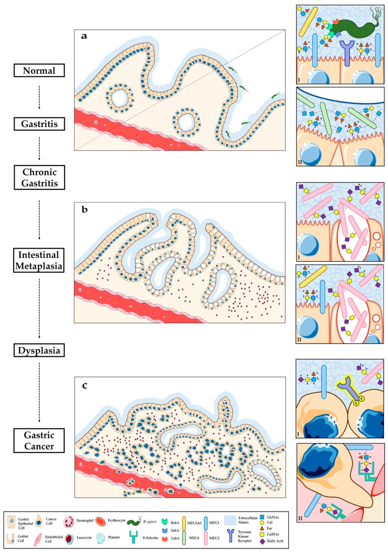
Figure 1.
Schematic representation of mucin expression pattern and associated O-glycan signatures through the gastric carcinogenesis cascade: (a) Normal mucous-secreting gastric epithelium (left) and early stage of H. pylori colonization (right); (aI) Superficial foveolar epithelial cells expressing membrane-bound MUC1 and secreted MUC5AC associated with type 1 Lewis antigens, which serve as receptors for H. pylori BabA-mediated adhesion, promoting infection and leading to gastritis; (aII) Glandular epithelial cells expressing the secreted MUC6 associated with type 2 Lewis antigens and the terminal α1,4-GlcNAc natural antibiotic glycan structure; (b) Intestinal metaplasia (IM) with goblet cells and inflammatory infiltrate; (bI) Complete type IM, with marked secretion of intestinal MUC2 carrying STn and absence of gastric mucins; (bII) Incomplete type IM, with co-expression of gastric and intestinal mucins; (c) Intestinal type gastric adenocarcinoma with disorganized glandular architecture and inflammatory infiltrate; (cI) Gastric cancer cells displaying aberrant surface glycosylation with concomitant activation of TKR-dependent signalling pathways; (cII) Sialylated Lewis antigens potentiate the invasive and metastatic capacity of GC cells, by serving as ligands to endothelial selectins during tumour cell extravasation.

