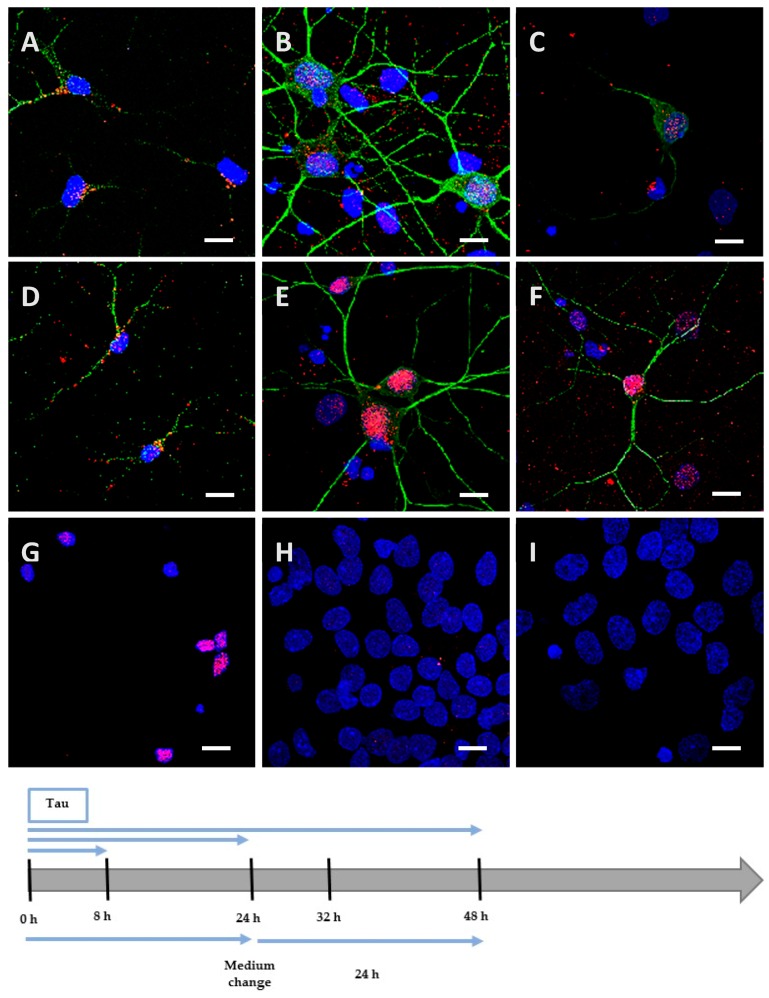
Figure 3.
Internalization and processing of the Tau protein (15 µg; A–C,H) and its highly phosphorylated form (1.5 µg; D–F,I) by cortical cells. Following 8 h (A,D), 24 h (B,E), or 48 h (C,F) of incubation with protein, cells were immunostained with a mouse anti-His-tag antibody targeted by an AlexaFluor® 594 antibody (red) and a rabbit anti-MAP2 antibody targeted by an AlexaFluor® 488 antibody (green). In other experiments, cells were placed in contact with protein for 24 h, whereupon the medium was replaced by one devoid of protein and maintained for an additional 24 h (H,I). The negative control (G) showed a non-specific distribution of the anti-His-tag, localized only in the nucleus. Nuclei were stained with DAPI. The protein was sometimes detected outside cells where it was probably attached to the extracellular matrix. All scale bars correspond to 10 µm.

