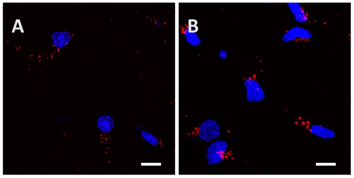
Figure A6.
Low phosphorylated (15 µg; A) and highly phosphorylated Tau proteins (1.5 µg; B) were visible inside cortical cells after 24 h of incubation even when a washing with 0.05% of trypsin for 1 min was performed. Trypsin was used to remove potential proteins attached to the extracellular matrix or the cytoplasmic membrane. Scale bars, 10 µm.

