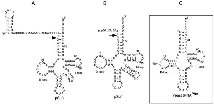
Figure 1.
The secondary structures of precursor tRNATyrSu3, precursor tRNASerSu1, and yeast tRNAPhe. (A,B) Precursor tRNATyrSu3 (pSu3) and tRNASerSu1 (pSu1). Black arrows indicate the canonical RNase P cleavage sites; (C) The grey arrow marks the major Pb2+-induced cleavage site in the D-loop of yeast tRNAPhe.

