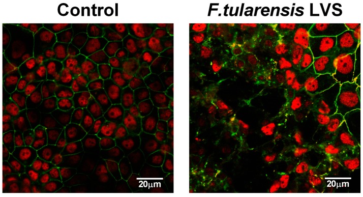
Figure 2.
Disruption of tight junctions in bronchial epithelial cells after F. tularensis infection. Polarised 16HBE cells were apically infected with F. tularensis LVS (MOI of 1) for 72 h and distribution of the tight junction protein occludin (green) analysed by confocal fluorescence microscopy. Nuclei stained with DAPI were shown in pseudo-colouring (red) representative image of three independent experiments.

