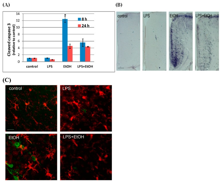
Figure 1.
LPS attenuates P7 ethanol-induced caspase-3 activation and morphological changes in microglia. (A) LPS (0.5g/kg)/saline was injected (i.p.) into P7 mice 2 h before ethanol (EtOH) (2.5g/kg, twice, 2 h apart)/saline injection, and 8 h and 24 h after the first ethanol injection, forebrains were taken and homogenates were analyzed by Western blots. The content of CC3 was normalized by actin and expressed as the ratio to the control. * Significantly different from all other groups by the Bonferroni post-hoc test after one-way ANOVA for 8 h samples; (B) P7 mice were treated as described in A, and 8 h after the first ethanol injection, mice were perfusion-fixed and brain sections were stained using anti-CC3 antibody. The representative images show the cingulate cortex region, and the bar indicates 200 µm; (C) Brain sections prepared as described in B were dual-labeled with anti-Iba1 (red) and anti-cleaved tau (green) antibodies. The bar indicates 20 µm.

