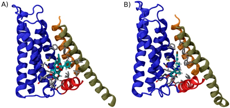
Figure 2.
Binding mode of TRPV1 modulators. Structure of TRPV1 in complex with: RTX (A); and capsazepine (B). For clarity only the structural element surrounding the vanilloid binding site are shown in cartoon representation using the same color code used in Figure 1. Amino acid side chains contacting the ligands are shown as sticks; to highlight the location of ligand in the two structures, the carbon atoms of RTX and capsezepine are highlighted by the cyan color. Note how the conformations of RTX and capsazepine are very similar, except for a phenyl group, which, in RTX, contacts the side chains of S6.

